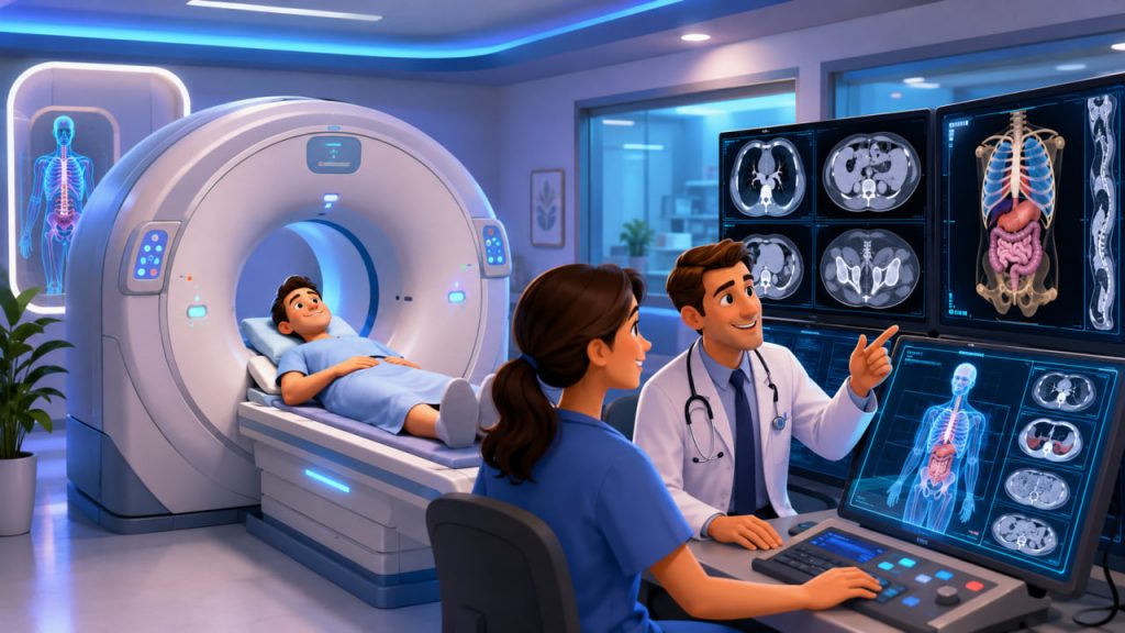
Computed tomography, commonly called a CT scan, is one of the most important diagnostic technologies in modern medicine. It can reveal internal injuries, tumors, infections, blood clots, kidney stones, bone damage, and many other conditions within minutes.
Unlike a conventional X-ray, which produces a flat image, CT combines multiple X-ray measurements to create detailed cross-sectional views of the body.
A CT scan provides much more anatomical detail than an ordinary radiograph, but it also usually involves a higher radiation dose.
What Is Computed Tomography?
Computed tomography is an imaging method that uses rotating X-ray equipment and computer processing to create thin images known as slices.
During the examination, an X-ray tube rotates around the patient. Detectors on the opposite side measure how much radiation passes through different tissues.
A computer reconstructs these measurements into cross-sectional images. The slices can be viewed individually, combined into three-dimensional models, or examined from different angles.
Bones, organs, blood vessels, fat, air, and abnormal tissue absorb X-rays differently, allowing the scanner to distinguish internal structures with considerable detail.
How Is CT Different From a Standard X-Ray?
A standard X-ray usually creates one or several two-dimensional images. Structures may overlap, making small abnormalities difficult to see.
A CT scanner collects many measurements from different directions. Computer reconstruction separates the body into virtual slices, reducing anatomical overlap.
For example, a chest X-ray may show a suspicious shadow, while a chest CT can help determine its exact location, shape, density, and relationship to nearby structures.
CT is therefore especially useful when doctors need a rapid and detailed view of internal anatomy.
What Conditions Can a CT Scan Detect?
Doctors use CT in emergency care, cancer medicine, neurology, cardiology, surgery, orthopedics, and many other specialties.
A CT scan may help identify:
- Internal bleeding
- Stroke and brain injury
- Bone fractures
- Lung disease
- Tumors and enlarged lymph nodes
- Kidney and urinary stones
- Appendicitis
- Bowel obstruction
- Blood clots
- Aneurysms
- Infections and abscesses
- Damage after an accident
CT can also help doctors plan surgery, radiotherapy, biopsies, drainage procedures, and other treatments.
The value of CT is particularly high in emergencies, where rapid diagnosis can directly affect survival and treatment decisions.
What Happens During a CT Scan?
The patient lies on a narrow table that moves through a large circular opening in the scanner.
Unlike a traditional MRI machine, a CT scanner usually has a short, open ring rather than a long tunnel. This can make the procedure easier for people who experience claustrophobia.
The radiologic technologist operates the equipment from a nearby control room and communicates through an intercom.
During the scan, the patient may be asked to:
- Remain still
- Raise the arms
- Hold the breath briefly
- Lie in a particular position
- Avoid eating beforehand when contrast is planned
The scanning itself may take only seconds, although preparation and contrast administration can make the appointment longer.
Movement can blur the images, so following breathing and positioning instructions is important.
What Is Contrast Material?
Some CT examinations use contrast material to make organs, blood vessels, or the digestive system easier to see.
Iodine-based contrast may be injected into a vein. Barium-based contrast may be swallowed or introduced through the digestive tract, depending on the examination.
After an intravenous injection, a patient may briefly experience:
- A warm sensation
- A metallic taste
- A feeling similar to urination
- Mild nausea
These sensations usually disappear quickly.
Contrast is not required for every CT scan. The decision depends on the body area and the clinical question.
Are Contrast Reactions Dangerous?
Most patients tolerate modern iodine-based contrast without serious problems.
Mild reactions may include itching, a rash, nausea, or sneezing. Severe allergic-like reactions are rare, and radiology departments are prepared to treat them.
Patients should tell the medical team about:
- Previous contrast reactions
- Kidney disease
- Diabetes
- Thyroid problems
- Asthma
- Current medications
- Pregnancy
- Possible dehydration
In some patients with reduced kidney function, intravenous contrast may worsen renal function. The medical team may review blood tests, modify the examination, or use another imaging method when necessary.
Does CT Use Radiation?
Yes. CT scanners use ionizing X-ray radiation.
Ionizing radiation contains enough energy to alter atoms and potentially damage cellular DNA. At the doses used in diagnostic imaging, the principal concern is a small possible increase in lifetime cancer risk rather than immediate radiation illness.
The dose varies widely according to:
- Body region
- Patient size
- Number of scan phases
- Scanner settings
- Clinical purpose
- Equipment technology
- Whether follow-up images are required
A CT examination generally involves more radiation than an ordinary X-ray because it collects many projections around the body.
How Dangerous Is a CT Scan?
For an individual medically justified examination, the radiation risk is usually small compared with the benefit of obtaining an accurate diagnosis.
The FDA identifies the major CT-related concerns as radiation exposure, possible contrast reactions, and incidental findings that may lead to additional investigations.
There is no universal safe maximum number of CT scans. The important question is whether each examination is clinically necessary and properly optimized.
Avoiding a necessary CT scan may be more dangerous than the radiation exposure if delayed imaging prevents doctors from detecting internal bleeding, stroke, cancer, or another serious condition.
Why Children Need Special Protection
Children are more sensitive to radiation because their tissues are developing and they have more years ahead during which a radiation-related effect could potentially develop.
The FDA recommends using child-sized scanning protocols and performing pediatric CT only when the expected information will contribute to diagnosis or treatment.
An adult scanning protocol should not automatically be used for a small child.
Doctors may also consider ultrasound or MRI when these methods can provide the necessary information without ionizing radiation.
CT Scans During Pregnancy
A pregnant patient, or someone who might be pregnant, should inform the medical team before the scan.
CT of the head, chest, arms, or legs usually results in little or no direct exposure to the fetus. Abdominal and pelvic CT requires more careful assessment because the fetus may be closer to the X-ray beam.
A necessary examination may still be performed when the expected benefit outweighs the potential risk. The protocol can often be modified to reduce exposure.
Doctors may choose ultrasound or MRI when these provide an appropriate alternative, but emergency CT should not automatically be delayed when it is needed to protect the mother’s health.
How Radiation Dose Is Reduced
Medical radiation protection follows two main principles: justification and optimization.
Justification means that the examination should answer an important medical question or change patient management.
Optimization means obtaining adequate diagnostic information using the lowest radiation dose reasonably necessary.
Modern scanners may reduce exposure through:
- Automatic exposure control
- Patient-sized protocols
- Limiting the scanned area
- Faster detector systems
- Advanced image reconstruction
- Avoiding unnecessary repeated phases
- Reviewing previous examinations
Very low-dose images are not always useful. If the dose is reduced too far, excessive image noise may hide disease and require the scan to be repeated.
CT, MRI, and Ultrasound: Which Is Better?
No imaging method is best for every problem.
CT is fast and excellent for trauma, bones, lungs, internal bleeding, and many emergency conditions.
MRI does not use ionizing radiation and often provides superior detail for the brain, spinal cord, joints, muscles, and certain soft tissues. However, it usually takes longer and may be unsuitable for some patients with metallic implants.
Ultrasound also avoids ionizing radiation. It is widely used for pregnancy, the abdomen, heart, blood vessels, and superficial tissues, but image quality can be limited by bone, air, body size, and anatomical location.
The best test is the one most likely to answer the clinical question accurately and promptly.
Expert Perspective
The World Health Organization emphasizes that medical imaging should be justified and optimized: the procedure must have a valid clinical purpose, and the dose should be kept as low as possible while still producing adequate diagnostic information.
The International Atomic Energy Agency takes the same approach, focusing on appropriate referral, patient-specific protocols, equipment quality control, and avoidance of unnecessary repeat scanning.
This expert guidance supports a balanced message:
CT should not be feared when it is medically necessary, but it should never be performed casually without a clear reason.
Preparing for a CT Examination
Follow the instructions provided by the imaging center.
Depending on the scan, you may need to avoid food for several hours, drink water, remove metal objects, or arrive early for contrast preparation.
Bring information about:
- Current medications
- Allergies
- Kidney problems
- Previous contrast reactions
- Pregnancy
- Earlier scans
- Implanted medical devices
Ask whether contrast will be used and when the written radiology report will be available.
Interesting Facts
- The word tomography refers to imaging the body in sections or slices.
- CT was previously widely called a CAT scan, meaning computed axial tomography.
- Modern scanners can create three-dimensional images from thin cross-sectional slices.
- CT can detect internal bleeding within minutes.
- No X-ray radiation remains inside the body after the scan ends.
- Contrast material does not produce radiation.
- CT angiography can examine arteries and veins after intravenous contrast injection.
- A scan may reveal an unrelated abnormality known as an incidental finding.
- Modern reconstruction software can produce useful images from lower-dose data.
- The radiation dose from CT varies considerably between different examinations.
Glossary
- Computed Tomography — An imaging method that combines X-rays and computer processing to create cross-sectional images.
- CT Scan — The examination performed using computed tomography equipment.
- Slice — A thin cross-sectional image representing one level of the body.
- X-Ray Tube — The rotating component that produces X-rays inside a CT scanner.
- Detector — A device that measures X-rays after they pass through the body.
- Contrast Material — A substance used to make organs, blood vessels, or tissues more visible.
- Iodinated Contrast — Iodine-containing contrast commonly injected during CT imaging.
- Ionizing Radiation — Radiation energetic enough to alter atoms and potentially damage cells.
- Radiation Dose — The amount of radiation delivered to or absorbed by the body.
- CT Angiography — CT imaging designed to examine blood vessels after contrast administration.
- Incidental Finding — An unexpected abnormality discovered while investigating another problem.
- Image Reconstruction — Computer processing that converts scanner measurements into medical images.
- Justification — Confirmation that an examination has a valid medical reason.
- Optimization — Adjustment of a scan to produce adequate images with the lowest necessary radiation dose.
- Radiologist — A physician specializing in interpreting medical images and performing image-guided procedures.