
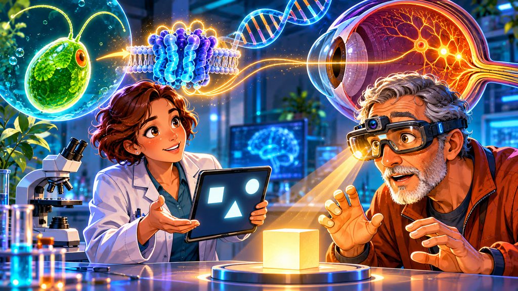
Imagine a tiny green alga swimming toward sunlight. Its ability to detect light might seem insignificant, yet studying this microscopic organism helped scientists develop a revolutionary way to control nerve cells. Decades later, that discovery contributed to an experimental treatment allowing people with severe blindness to detect objects again. In October 2026, the scientific breakthroughs behind this technology received the Nobel Prize in Physiology or Medicine.
The discovery connects marine biology, genetics, neuroscience, and ophthalmology in an extraordinary story. A light-sensitive protein originally found in algae became a molecular switch for nerve cells. Researchers eventually adapted this mechanism to help people affected by inherited retinal diseases. Although the treatment has not restored normal sight, its results represent an important advance in efforts to recover lost vision.
Who Won the 2026 Nobel Prize in Medicine?
On October 5, 2026, the Nobel Assembly announced that Peter Hegemann, Georg Nagel, and Karl Deisseroth would share the Nobel Prize in Physiology or Medicine. The scientists were recognized for discoveries that established optogenetics, a technology that uses light to control selected living cells. Their work transformed how researchers investigate the nervous system and helped create new possibilities for treating neurological diseases. One particularly remarkable application involves restoring limited visual function to people who have lost the ability to see.
The Nobel Prize honored the foundational discoveries behind optogenetics, rather than a specific treatment for blindness. Hegemann and Nagel identified a remarkable light-sensitive protein in algae, while Deisseroth helped transform the discovery into a practical method for controlling neurons. Their combined achievements provided the foundation for later clinical research. A discovery made while investigating microscopic algae ultimately opened an entirely new direction in medicine.
How Did Scientists Discover the Light-Sensitive Algae Protein?
The story began with Chlamydomonas, a single-celled green alga capable of swimming toward light. Scientists wanted to understand how such a simple organism could detect illumination and respond almost instantly. Research revealed proteins called channelrhodopsins, which act as molecular gates in cell membranes. When exposed to certain wavelengths of light, these proteins allow electrically charged particles to enter cells.
This mechanism is known as a light-gated ion channel. The movement of charged particles changes the electrical state of the cell, creating a signal that can influence its activity. Researchers discovered that the proteins could also function when introduced into other types of cells. This unexpected versatility made channelrhodopsins especially valuable for neuroscience.
How Can an Algae Protein Control Human Nerve Cells?
Human nerve cells communicate through electrical impulses, allowing the brain to process information and coordinate the body. By introducing genetic instructions for channelrhodopsins into selected neurons, scientists can make those cells respond to controlled flashes of light. This is the essential principle of optogenetics, a technique combining genetic engineering with precise optical stimulation. It allows researchers to investigate how specific groups of neurons contribute to memory, movement, and behavior.
In 2005, Karl Deisseroth and colleagues demonstrated that light-sensitive proteins could activate neurons using flashes of blue light. Subsequent experiments extended the technique to neural circuits in living animals. Unlike conventional electrical stimulation, optogenetics can target genetically defined populations of cells. The discovery suggested that light might eventually help compensate for damaged biological systems, including parts of the human eye.
Why Do Some People Lose Their Vision?
The human eye contains a specialized layer of nerve tissue called the retina. Within it, photoreceptors known as rods and cones convert incoming light into electrical signals. Other retinal neurons process those signals before transmitting information toward the brain. When photoreceptors deteriorate, the normal process of vision becomes increasingly difficult.
One major cause of inherited blindness is retinitis pigmentosa, a group of genetic diseases that progressively damage the retina. Symptoms commonly begin with difficulty seeing at night, followed by narrowing peripheral vision and, in severe cases, profound visual loss. However, some nerve cells can survive even after the photoreceptors have disappeared. This gave researchers an opportunity to bypass the damaged cells rather than replace the entire retina.
The 2021 Breakthrough: A Blind Man Regained Partial Sight
In 2021, an international team led by ophthalmologist José-Alain Sahel and neuroscientist Botond Roska reported an extraordinary clinical result. Their patient was a 58-year-old man diagnosed with retinitis pigmentosa approximately 40 years earlier. Before treatment, his remaining vision was largely limited to detecting light. After experimental therapy and visual training, he regained the ability to perceive, locate, count, and touch certain objects.
During testing, the man could identify the positions of objects placed on a table while wearing specialized goggles. He also reported detecting the white stripes of a pedestrian crossing. These achievements were especially significant because the underlying retinal disease had already caused extensive damage. The treatment did not restore ordinary eyesight, but it demonstrated that a damaged visual system could recover some useful function.
How Does the Experimental Vision Restoration Treatment Work?
Researchers used a modified viral vector to deliver genetic instructions for a protein called ChrimsonR into the patient’s eye. This engineered protein belongs to the channelrhodopsin family and originates from research involving the green alga Chlamydomonas noctigama. It responds to orange-red light, making it suitable for a different stimulation system from the blue-light-sensitive proteins used in early optogenetic experiments. The treatment targets surviving retinal ganglion cells, giving them an artificial ability to respond to light.
Special goggles provide the second essential component of the treatment. A built-in camera captures the surroundings, while a processor converts visual information into simplified patterns. A tiny projector directs controlled amber-colored light pulses onto the treated retina, activating cells containing ChrimsonR. The brain then receives signals that can help the patient identify the presence and position of objects.
The New 2026 Study: What Happened to Ten Patients?
On October 7, 2026, researchers published additional clinical results in the New England Journal of Medicine. The study involved ten people with advanced retinitis pigmentosa who received experimental optogenetic therapy. Seven participants demonstrated improved light sensitivity, and six achieved improvements considered clinically meaningful by the study’s criteria. Four participants showed consistent progress across multiple functional vision tests over repeated assessments.
Some patients became better able to detect objects, determine their locations, and reach toward them using the specialized goggles. Most eye-related adverse events were mild or moderate, including temporary inflammation and increased eye pressure. The results extended the earlier single-patient breakthrough to a larger group. Nevertheless, this was still a small, early-stage clinical trial, and the treatment did not restore normal vision, reading ability, or facial recognition.
Why Is Restoring Normal Vision Still So Difficult?
Natural vision is extraordinarily sophisticated. Healthy retinas contain millions of photoreceptors and complicated networks that process contrast, color, movement, and spatial details. The experimental system cannot reproduce all these functions simply by making surviving neurons light-sensitive. Instead, it delivers simplified information that the brain must learn to interpret.
Researchers also need to improve image resolution, light sensitivity, treatment consistency, and long-term safety. Different patients may have different amounts of surviving retinal tissue, influencing treatment outcomes. Visual training is another important part of adapting to the new signals. The central challenge is no longer merely generating an electrical response to light, but transforming that response into increasingly useful visual perception.
Could Optogenetics Transform Medicine Beyond Blindness?
Optogenetics has already revolutionized laboratory studies of the brain by allowing researchers to activate or inhibit particular neural circuits. Scientists use it to investigate mechanisms involved in movement, learning, memory, and neurological disorders. Potential medical applications extend beyond retinal disease, although many remain experimental. The technique offers an unusually precise way to examine relationships between nerve-cell activity and biological function.
For vision restoration, future developments may involve improved light-sensitive proteins, more advanced image processors, or targeting different surviving retinal cells. Treatments might eventually help people affected by other retinal disorders, depending on which neural structures remain functional. As of November 2026, ChrimsonR-based vision restoration remains experimental rather than an established clinical cure. Its greatest achievement so far is demonstrating that limited visual function can return even after substantial photoreceptor loss.
A tiny organism that once attracted the curiosity of biologists has helped create an entirely new medical technology. The journey from algae to Nobel Prize-winning neuroscience shows how unexpected discoveries can reshape our understanding of human biology. Which future application of optogenetics interests you most: restoring vision, treating neurological diseases, or understanding how the brain works? Share your thoughts in the comments.
Interesting Facts
- The 2026 Nobel Prize in Physiology or Medicine was awarded to Peter Hegemann, Georg Nagel, and Karl Deisseroth.
- The discovery began with research into how single-celled green algae respond to light.
- Channelrhodopsins can make previously light-insensitive nerve cells respond to illumination.
- The first documented case of partial human vision restoration using optogenetics was published in 2021.
- The original patient was 58 years old and had been diagnosed with retinal disease around four decades earlier.
- In a 2026 study, seven of ten participants showed improved sensitivity to light.
- Experimental vision restoration requires genetic modification of retinal cells combined with specialized light-stimulating goggles.
Glossary
- Optogenetics — A technique that combines genetic engineering and light to control the activity of selected cells.
- Channelrhodopsins — Light-sensitive proteins originally identified in algae that can act as ion channels.
- Light-Gated Ion Channel — A membrane protein that opens in response to light, allowing electrically charged particles to move across a cell membrane.
- Retina — The light-sensitive layer of nerve tissue located at the back of the eye.
- Photoreceptors — Specialized retinal cells that detect light, including rods and cones.
- Retinitis Pigmentosa — A group of inherited disorders that progressively damage retinal photoreceptors.
- Viral Vector — A modified virus used to deliver genetic material into targeted cells.
- ChrimsonR — An engineered light-sensitive protein used in experimental optogenetic vision restoration.
- Retinal Ganglion Cells — Nerve cells that transmit visual information from the retina toward the brain.