Fluorography is a chest-imaging method traditionally used for large-scale screening of tuberculosis and other abnormalities in the lungs and surrounding structures. It is especially familiar in countries where adults have historically undergone regular preventive chest examinations.
The term can be confusing because it resembles fluoroscopy, a different imaging procedure used to observe movement inside the body in real time. In modern healthcare, traditional film fluorography has largely been replaced by digital chest radiography, but the older name is still widely used.
Fluorography is primarily a screening examination. An abnormal image does not establish a final diagnosis and usually requires additional medical evaluation.
What Is Fluorography?
Traditional photofluorography records a small image of the chest from an X-ray fluorescent screen. It was developed as an economical way to examine large numbers of people, particularly during tuberculosis-control campaigns.
Instead of producing a full-size radiograph directly, older systems photographed the image displayed on a fluorescent screen onto small-format film.
Modern digital equipment works differently. It uses an electronic detector to capture the X-ray image and display it on a computer. Digital imaging generally provides better processing, easier storage, faster transmission, and more flexible viewing than small-film fluorography.
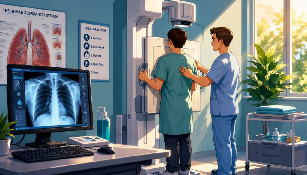
For patients, the basic experience is similar to an ordinary chest X-ray: the person stands against a detector while the equipment produces an image of the chest.
How Does the Examination Work?
X-rays pass through the body at different rates.
Air-filled lungs allow more radiation to pass through and normally appear relatively dark. Dense tissues such as bones absorb more radiation and appear lighter. The heart, blood vessels, muscles, and other structures create intermediate shades.
During a standard examination, the patient is usually asked to:
- Remove metal objects and clothing that may cover the chest
- Stand facing the detector
- Position the shoulders and arms correctly
- Take a deep breath
- Hold the breath for a few seconds
- Remain completely still during exposure
The actual exposure takes only a fraction of a second. The entire appointment is normally brief unless extra images are required.
Holding the breath expands the lungs and reduces motion, helping the medical professional obtain a clearer image.
What Can Fluorography Show?
Chest imaging can reveal visible changes in the lungs, heart, ribs, diaphragm, and surrounding tissues.
Possible findings include:
- Areas of inflammation
- Lung infiltrates
- Cavities
- Fluid around the lungs
- Scarring
- Certain tumors or nodules
- Changes associated with tuberculosis
- Enlargement of the heart
- Some skeletal abnormalities
However, many conditions can produce similar appearances.
For example, a shadow may represent an old scar, active infection, technical artifact, overlapping anatomy, or another process. Further investigation may be necessary to determine its cause.
Fluorography can identify a suspicious abnormality, but it cannot reliably explain every abnormality by itself.
Fluorography and Tuberculosis Screening
Fluorography became closely associated with tuberculosis because pulmonary tuberculosis often produces visible changes on chest images.
Chest radiography remains an important tool for identifying people who may require further testing. The World Health Organization describes chest radiography as valuable for the early detection and screening of pulmonary tuberculosis.
Nevertheless, an X-ray image alone cannot confirm tuberculosis.
A person with suspicious findings may need:
- Sputum testing
- Molecular diagnostic testing
- Microscopy or culture
- Clinical assessment
- Additional imaging
- Evaluation by a tuberculosis specialist
Chest radiography is relatively sensitive for detecting possible pulmonary tuberculosis, but its specificity is limited. This means it may reveal many abnormalities that are not caused by tuberculosis.
Who May Benefit From Screening?
Modern medical screening should be based on risk rather than performed automatically without considering individual circumstances.
WHO recommendations prioritize systematic tuberculosis screening for groups in whom the potential benefits are likely to outweigh the harms. These may include close contacts of people with tuberculosis, people living with HIV, individuals exposed to silica dust, prisoners, and other populations with an increased risk of infection or delayed diagnosis.
The need for routine chest imaging depends on:
- Local tuberculosis prevalence
- Occupational exposure
- Known contact with tuberculosis
- Symptoms
- Immune status
- Age
- Previous medical history
- National health regulations
A healthy person at low risk may not benefit from frequent repeated imaging. Screening schedules should therefore be determined by local medical guidance rather than habit alone.
Symptoms That Require Medical Attention
A normal recent fluorography result does not mean that future symptoms should be ignored.
A person should seek medical evaluation for symptoms such as:
- A persistent cough
- Coughing up blood
- Unexplained weight loss
- Fever
- Night sweats
- Shortness of breath
- Persistent chest pain
- Unusual weakness
Some diseases may not be visible during their earliest stages. Others can develop after the examination.
Screening does not replace a medical consultation when symptoms are present.
Fluorography, Chest X-Ray, and Fluoroscopy
These terms are related but not identical.
Traditional fluorography usually refers to miniature chest imaging developed for mass screening.
A chest X-ray, or chest radiograph, produces a diagnostic image of the chest. Modern digital radiography now performs many functions historically associated with fluorography.
Fluoroscopy is a separate technique that creates a continuous or rapidly updated X-ray image. Doctors use it to observe movement, guide instruments, study swallowing, or examine the digestive and cardiovascular systems.
Fluoroscopy may involve a longer radiation exposure than a single chest image because it records a moving process rather than one brief exposure.
Radiation Exposure and Safety
Fluorography and chest radiography use ionizing radiation.
Modern chest X-ray doses are generally low. RadiologyInfo estimates the typical effective dose from an adult chest X-ray at approximately 0.1 millisievert, comparable to about ten days of natural background radiation. The actual dose varies according to the equipment, number of views, patient size, and imaging technique.
A low dose does not mean zero risk. Medical imaging should still follow the principle that exposure should be justified and kept as low as reasonably achievable.
Patients should inform medical staff if they are pregnant or may be pregnant. The healthcare professional can determine whether the examination is necessary, whether it can be postponed, or whether another approach is appropriate.
The potential benefit of a medically justified examination usually outweighs the very small radiation risk.
What Happens After an Abnormal Result?
An abnormal fluorography result does not necessarily mean that a serious disease is present.
The next step may involve comparison with previous images. Stable findings that have remained unchanged for years may be less concerning than a newly developed abnormality.
The doctor may recommend:
- A full diagnostic chest radiograph
- Computed tomography
- Laboratory tests
- Sputum analysis
- Pulmonary-function testing
- Consultation with a pulmonologist
- Follow-up imaging after a defined period
The correct next step depends on the appearance, symptoms, age, medical history, and risk factors.
Patients should not attempt to interpret the image without professional assistance. Small technical details can substantially affect radiological interpretation.
Advantages of Digital Chest Imaging
Digital systems have transformed chest screening.
Images can be enlarged, adjusted, compared with earlier examinations, stored electronically, and transmitted to another specialist. This can improve access to radiological interpretation in regions with limited local expertise.
Digital equipment also supports computer-aided detection. WHO guidance includes the use of chest radiography and, in appropriate screening programs, computer-assisted systems that identify images requiring further review.
Artificial intelligence may help prioritize suspicious examinations, but it does not remove the need for clinical judgment or confirmatory testing.
Expert Perspective
The World Health Organization emphasizes that chest radiography is particularly useful as a screening and triage tool for tuberculosis rather than a stand-alone diagnostic test. An abnormal image should lead to appropriate clinical and laboratory evaluation.
This distinction is essential. Screening aims to identify people who may have a disease, while diagnosis aims to determine whether they actually have it.
Fluorography is most valuable when it forms part of a complete medical pathway rather than being treated as a final answer.
Interesting Facts
- Traditional photofluorography photographed an image from a fluorescent X-ray screen.
- The technique was designed to make mass chest screening faster and less expensive.
- Modern digital radiography has replaced small-film fluorography in many healthcare systems.
- The lungs appear relatively dark on an X-ray because they contain air.
- Metal jewelry, buttons, and clothing decorations may create misleading shadows.
- A chest image may reveal abnormalities outside the lungs, including changes in the heart or bones.
- Chest radiography can suggest tuberculosis but cannot confirm it without additional testing.
- Artificial intelligence can help identify suspicious chest images in large screening programs.
- The radiation dose from a modern chest X-ray is usually much lower than that from a chest CT scan.
- Fluorography and fluoroscopy are different procedures despite their similar names.
Glossary
- Fluorography — A chest-imaging technique historically used for large-scale X-ray screening.
- Photofluorography — The traditional process of photographing an X-ray image from a fluorescent screen.
- Chest Radiography — X-ray imaging used to examine the lungs, heart, ribs, and nearby structures.
- Fluoroscopy — Real-time or moving X-ray imaging used to observe internal processes or guide procedures.
- Tuberculosis — An infectious disease caused by bacteria of the Mycobacterium tuberculosis group.
- Screening — Testing people who may not have symptoms to identify possible disease.
- Diagnosis — The process of determining which disease or condition is present.
- Infiltrate — An area of abnormal material or increased density visible in lung tissue.
- Pulmonary Nodule — A small rounded opacity in the lung that may have many possible causes.
- Ionizing Radiation — Radiation with enough energy to alter atoms and potentially damage biological tissue.
- Millisievert — A unit used to describe effective radiation dose.
- Digital Detector — An electronic device that captures an X-ray image.
- Computer-Aided Detection — Software that helps identify potentially abnormal features in medical images.
- Triage Test — An initial test used to decide who requires more detailed diagnostic evaluation.