A PET scan, short for Positron Emission Tomography, is a highly advanced medical imaging technique that allows doctors to observe how organs and tissues function in real time. Unlike traditional X-rays or CT scans, which show the structure of the body, PET scans reveal the activity occurring inside — such as metabolism, blood flow, and cellular behavior. This makes PET one of the most powerful tools for diagnosing cancer, neurological disorders, and heart disease. PET imaging combines radiochemistry, physics, and computer science to create detailed maps of biochemical processes, helping specialists detect disease early and monitor treatment progress.
PET scans work by using tiny amounts of radioactive tracers, often forms of glucose or other biologically active molecules. Because active cells absorb these tracers differently, PET scans can visualize how tissues are functioning at the molecular level. Cancer cells, for example, absorb more glucose than normal cells, making tumors appear brighter on PET images.
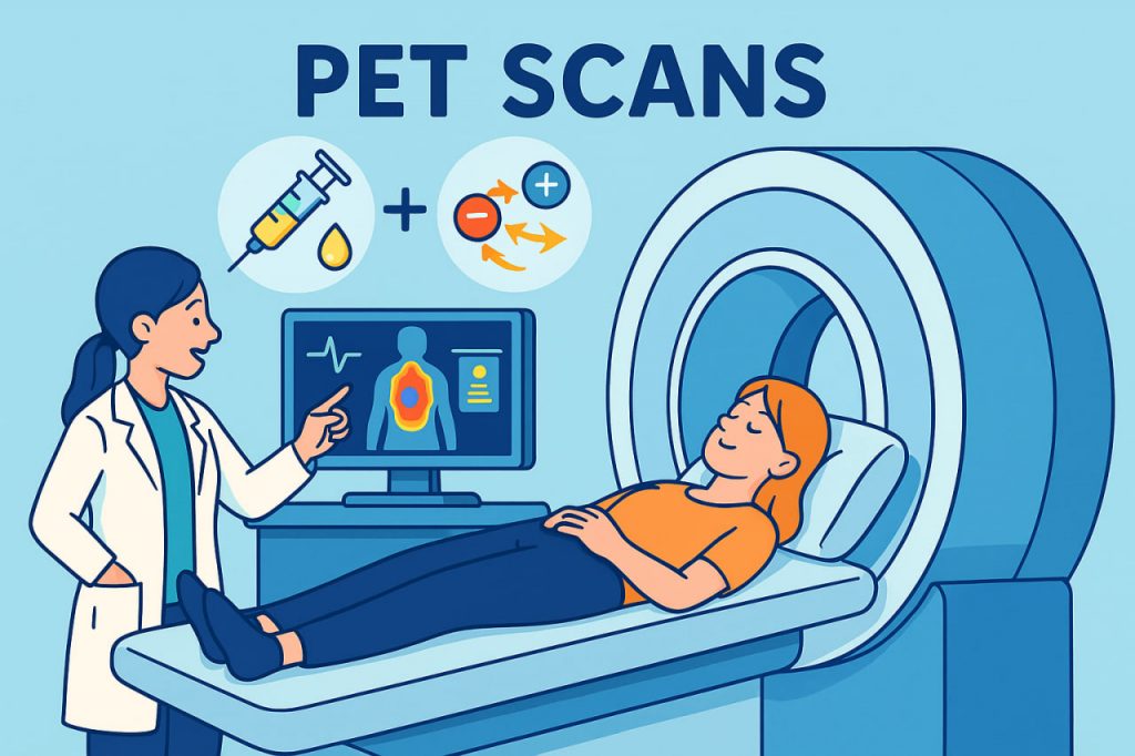
How a PET Scan Works
A PET scan involves several steps:
- Tracer Injection
A small amount of a radioactive molecule — called a radiotracer — is injected into the bloodstream. - Tracer Uptake
Organs and tissues absorb the tracer at different rates depending on their activity. - Particle Emission
The tracer emits positrons, the antimatter counterparts of electrons. - Annihilation Event
When a positron meets an electron, they annihilate and produce two gamma rays moving in opposite directions. - Detection & Image Formation
The PET scanner detects these gamma rays, and a computer reconstructs a detailed image showing biological activity inside the body.
According to medical physicist Dr. Clara Hammond:
“PET scans let doctors visualize disease before it changes anatomy —
they reveal function long before structure is affected.”
This ability to detect early biochemical changes is why PET scans are so important.
What PET Scans Are Used For
PET scans are invaluable across many fields of medicine:
Cancer Diagnosis & Monitoring
- detect early-stage tumors
- determine whether cancer has spread
- evaluate how well treatments are working
- distinguish between scar tissue and active cancer
Cancer cells often show bright “hot spots” on PET images due to high metabolic activity.
Neurology
PET scans help diagnose and study:
- Alzheimer’s disease
- epilepsy
- Parkinson’s disease
- brain injuries
- neurotransmitter disorders
They show areas of the brain that are overactive or underactive, offering insights unavailable through MRI alone.
Cardiology
PET scans reveal:
- blood flow to the heart
- areas of reduced metabolism
- early signs of coronary artery disease
This helps cardiologists determine whether parts of the heart muscle are still alive after an injury.
Types of Radiotracers
Different tracers highlight different biological processes:
- FDG (Fluorodeoxyglucose) — glucose metabolism
- Oxygen-15 — blood flow
- Nitrogen-13 — heart function
- Carbon-11 tracers — brain chemistry
- Amyloid tracers — Alzheimer’s disease detection
Each tracer provides unique diagnostic information.
Benefits of PET Scans
PET scans offer several key advantages:
- detect disease earlier than structural imaging
- provide functional and metabolic insights
- help tailor treatments
- allow precise monitoring over time
- safe when used appropriately
The radiation dose is relatively low and carefully controlled.
Limitations
Despite their strengths, PET scans have constraints:
- high cost of equipment and radiotracers
- tracers have short lifespans and require nearby production facilities
- lower spatial resolution than MRI
- require experienced specialists to interpret
PET scans are often combined with CT or MRI to provide both structural and functional detail.
Interesting Facts
- PET scans use positrons — the antimatter version of electrons.
- The most common PET tracer, FDG, behaves like glucose, revealing metabolic activity.
- PET scanners detect gamma rays produced inside the body during annihilation events.
- PET combined with CT or MRI provides some of the most detailed diagnostic images in modern medicine.
- PET technology originated from research in nuclear physics and particle detectors.
Glossary
- Radiotracer — a radioactive molecule injected into the body for imaging.
- Positron — the antimatter counterpart of the electron.
- Gamma Ray — a high-energy photon produced during annihilation events.
- Metabolic Activity — how active cells are in consuming energy.
- PET/CT — a combined scan that overlays metabolic and structural images.