Ultrasound (US), also known as sonography, is a medical imaging technique that uses high-frequency sound waves to create pictures of the inside of the body. Unlike X-rays or CT scans, ultrasound does not use ionizing radiation, making it one of the safest diagnostic tools available. It is widely used to examine soft tissues, blood vessels, and internal organs, as well as to monitor fetal development during pregnancy.
How Ultrasound Works
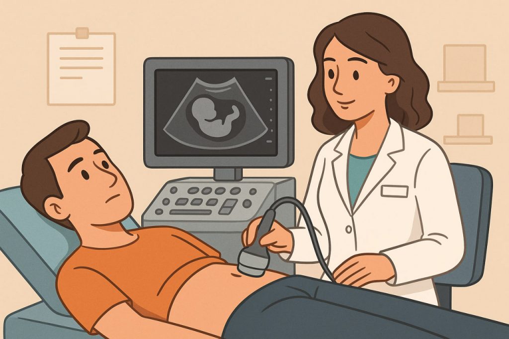
An ultrasound machine sends sound waves into the body through a handheld device called a transducer. These waves bounce off tissues, fluids, and organs, and the echoes are captured by the transducer. A computer then converts the echoes into real-time images that doctors can interpret. Because it is non-invasive and painless, ultrasound is often the first step in diagnosing various conditions.
Medical Uses of Ultrasound
Ultrasound is used in many areas of medicine, including:
- Pregnancy – monitoring fetal growth, detecting abnormalities, and checking placenta and amniotic fluid.
- Cardiology – echocardiograms evaluate the structure and function of the heart.
- Abdominal exams – to assess the liver, gallbladder, pancreas, kidneys, and bladder.
- Vascular health – detecting blood clots, blockages, or arterial narrowing.
- Musculoskeletal system – diagnosing tendon or ligament injuries.
Advantages of Ultrasound
Ultrasound is safe, affordable, and widely accessible. It does not expose the body to harmful radiation and provides real-time imaging. This makes it especially useful for guiding medical procedures, such as biopsies, or monitoring conditions over time.
How Often Should Ultrasound Be Done?
The frequency of ultrasound depends on the medical need:
- Pregnancy – typically 2–3 standard ultrasounds are recommended during a healthy pregnancy, unless the doctor advises more frequent scans.
- Chronic conditions – patients with liver, kidney, or heart issues may require regular ultrasounds for monitoring, as directed by a physician.
- Preventive purposes – routine ultrasounds are not recommended for healthy individuals without symptoms, since unnecessary imaging provides no additional health benefits.
It is important to note that ultrasound should always be performed based on a doctor’s recommendation, not as self-prescribed screening.
Conclusion
Ultrasound is a safe, non-invasive imaging method that provides valuable insights into the body’s internal structures. It plays a critical role in pregnancy monitoring, disease diagnosis, and treatment guidance. While it can be repeated without harm, it should be done only when medically justified. Consulting a healthcare professional ensures that ultrasound examinations are used effectively and appropriately.
Glossary
- Ultrasound (US) – an imaging technique that uses sound waves to create pictures of internal organs and tissues.
- Transducer – a handheld device that emits sound waves and receives echoes during an ultrasound scan.
- Sonography – another term for ultrasound imaging.
- Echocardiogram – an ultrasound of the heart used to evaluate its function and structure.
- Non-invasive – a medical procedure that does not require entry into the body or breaking the skin.