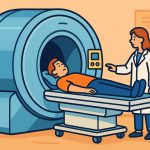
An X-ray machine is a medical device that uses ionizing radiation to create images of the inside of the human body. It is one of the most important diagnostic tools in modern medicine, allowing doctors to see bones, tissues, and certain organs without the need for surgery. X-rays have been used since their discovery in 1895 by Wilhelm Röntgen, and their applications have expanded greatly since then.
How X-Rays Are Produced
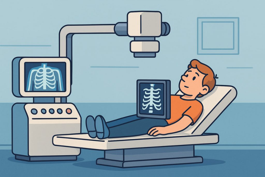
X-rays are a form of electromagnetic radiation with very short wavelengths and high energy. In an X-ray machine, they are produced when high-speed electrons collide with a metal target, usually made of tungsten. The sudden deceleration of electrons generates X-rays, which are then directed toward the body part being examined.
How Images Are Created
When X-rays pass through the body, different tissues absorb them at different rates. Dense materials like bones absorb more X-rays and appear white on the image. Softer tissues, such as muscles and fat, absorb less radiation and appear in shades of gray. Air-filled areas, like the lungs, appear darkest. These contrasts create a clear image that helps doctors detect fractures, infections, or abnormalities.
Uses of X-Ray Machines
X-ray machines are widely used in medicine:
- Bone fractures – quickly identifying broken or dislocated bones.
- Dental care – checking cavities, root conditions, and jaw structure.
- Chest scans – detecting lung infections, pneumonia, or tuberculosis.
- Mammography – screening for breast cancer.
- CT scans – combining multiple X-ray images to create 3D views of organs.
Outside medicine, X-ray technology is also used in airport security scanners, industrial inspection, and scientific research.
Safety and Risks
Because X-rays are a type of ionizing radiation, they can damage cells and DNA with excessive exposure. However, modern X-ray machines use very low doses, and the benefits usually outweigh the risks. Protective measures, such as lead aprons and shielding, are used to minimize exposure. Patients are only exposed for fractions of a second during scans, and repeated imaging is carefully managed by doctors.
Advances in X-Ray Technology
Modern X-ray machines are far safer and more precise than older models. Digital X-rays reduce radiation doses and provide faster results. Portable X-ray devices allow imaging outside hospitals, useful in emergencies or remote areas. As technology advances, X-ray machines continue to evolve, offering higher clarity and safer diagnostics.
Conclusion
X-ray machines revolutionized medicine by making it possible to look inside the human body without surgery. By using controlled doses of ionizing radiation, they help diagnose fractures, infections, and diseases quickly and effectively. With modern safety measures and digital improvements, X-ray technology remains one of the most valuable tools in healthcare and beyond.
Glossary
- X-rays – high-energy electromagnetic waves used for imaging.
- Electromagnetic Radiation – energy that travels as waves, including light, radio waves, and X-rays.
- Ionizing Radiation – radiation with enough energy to remove electrons from atoms and damage tissue.
- Tungsten Target – a metal surface used in X-ray machines to produce X-rays.
- CT Scan (Computed Tomography) – a technique that combines multiple X-ray images into a 3D model.
- Lead Apron – protective clothing that shields patients from unnecessary radiation.